


The ankle joint is often injured because of the heavy load it puts on. Diagnosis such as ankle arthrosis is not uncommon. It is placed regardless of the age and gender of the patient. What is ankle arthrosis and how can it be treated?
What is this?
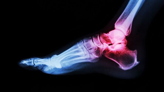
There is an incredible load on the ankle. Its function is to keep the body upright. Thanks to him, someone walked and ran. With a violation of the ankle system, it is very difficult to lead a normal lifestyle. What interferes with ankle work?
Ankle arthrosis, what is it? This is a chronic joint disease, characterized by degenerative processes. In the cartilage of the joints, an irreversible process is triggered, which leads to great complications.
Ankle arthrosis develops gradually. The surface of a healthy joint is elastic and smooth. They provide cushioning under heavy loads and glide smoothly while driving. With pathology, tissue trophism and metabolism are disturbed. The joint surface becomes inelastic and rough. During movement, the cartilage comes in contact with each other, which causes inflammation. During weight lifting, the main load falls on the bones, which threaten with degenerative disorders.
Lack of treatment leads to more serious disorders. At stage 3-4, damage to cartilage and tissues is observed. The synovium becomes inflamed. The joints become unstable. Support function violated. All these violations as a whole lead to the fact that movement becomes impossible.
Arthrosis (osteoarthritis) is one of the most common joint diseases that affects a large number of people.
Causes and risk factors
What is arthrosis of the ankle joint, we have solved it. Now let's find out what the main cause is. Ankle arthrosis is considered a pathology in old age. This is due to age -related changes in the body. Cartilage becomes thinner, bones become unstable and brittle. However, in the past decade, the diagnosis of ankle arthrosis has become much younger. Such statistics are disappointing, as many patients ignore the first signs of the disease. Late diagnosis always threatens the development of serious complications.
Provoking factors include:
- dislocation;
- bruises;
- inflammatory diseases;
- injuries;
- overweight;
- impaired metabolism;
- unbearable physical activity;
- wearing uncomfortable shoes;
- autoimmune and endocrine diseases;
- osteochondrosis.
Clinical symptoms
Ankle arthrosis is known by the following features:
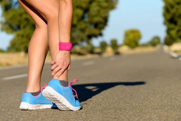
- It hurts. It is mild at first and appears after walking or doing physical activity. Sometimes a person is in an uncomfortable position. With the development of pathological processes, the pain syndrome increases and the worry is already resting.
- Swelling and inflammation. These signs appear against a background of injuries and dislocations. Body temperature in the affected area increases.
- Click. When the ankle is affected, click "dry" and cause an attack of pain.
- Dislocation or subluxation. Due to the thinning and shrinkage of cartilage tissue, the joints become unstable. Bones can slide and fall from the joint capsule. These changes cause severe pain.
- Joint stiffness. When cartilage tissue is replaced, the bone joint stops functioning normally, which negatively affects its movement.
- Joint deformities. Symptoms appear in stage 3-4 arthrosis. Osteophytes also cause curvature of the ankle.
If any of the symptoms appear, it is recommended to see a doctor immediately. Treatment started on time is a step towards recovery.
Arthrosis of the foot and ankle joints is characterized by slow progression with the gradual development of clinical manifestations over several years.
Classification and ranking
The disease develops in a variety of ways. In some patients, several years pass from the first symptoms to the final stage, in others, rapid progression of the disease is observed. The speed depends on the age and state of health of the patient, the time of initiation of therapy. The symptoms of arthrosis of the ankle joint become more pronounced with the progression of the disease.
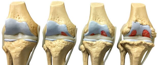
There are four stages of arthrosis:
- The first stage is often unnoticed. Sometimes morning cramps and ankle pain appear after strenuous exercise. As the legs move, the sound of cramps is heard. Pathological changes have not yet been seen on x-rays, but the process of cartilage destruction has already begun.
- Morning stiffness becomes prolonged. It takes 20-30 minutes to expand the legs. Sometimes lameness occurs. Grade 2 arthrosis of the ankle joint is recognized on roentgenogram by bone tissue growth, bone displacement.
- Symptoms at 3 levels are pronounced. The pain is no longer only after a heavy load, but also during rest. It is difficult for patients to do without painkillers. Lameness increased. Crutch disease may be required. The affected joints are swollen and deformed. Ankle muscle atrophy. X-rays show narrowing of the joint space, osteophyte formation, subluxation.
- Level 4 is the hardest. It develops due to lack of treatment. The cartilage is destroyed, the joint surface is united. Walking is no longer possible.
With the development of osteoarthritis of the ankle, there are gradual changes in the cartilage and bone tissue on the articular surface.
Diagnostics
The diagnosis of ankle arthrosis is based on clinical symptoms and information obtained during the examination. Laboratory studies are considered ineffective, as there are no special tests that can detect pathology. During the remission period, all indicators are within normal limits, with disease progression, clinical blood tests will show high levels of C-reactive protein and ESR. This indication indicates that the pathological process has already begun.
To confirm the diagnosis, instrumental methods are used:
- radiography;
- Magnetic resonance imaging;
- Ultrasound;
- bone scintigraphy;
- diagnostic joint puncture.
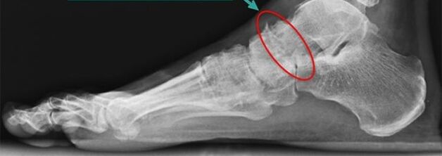
Brief radiography
Simple radiography is the most reliable and effective method for diagnosing diseases occurring in the musculoskeletal system. The principle of manipulation is the absorption of different X-rays by muscle tissue. Soft tissue allows X-rays to pass through, but hard tissue absorbs. X-rays allow you to diagnose the disease itself and its consequences.
Conventional radiography is a method of examination in which a small number of X-rays are sent through the body or body part of the person.
The image allows you to see:
- The state of the bone surface in articulation.
- The shape, size and arrangement of the structures at the joints are interrelated with each other.
- The condition of the fabric.
- The size of the joint space.
These indicators help doctors determine the type and degree of joint damage. If the data are insufficient, then the doctor prescribes another study.
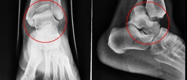
With ankle arthrosis, X-rays are performed in three projections:
- side;
- back;
- back with feet moved inwards.
The disease is characterized by the following changes:
- reduction of joint space;
- the presence of osteophytes;
- bone cartilage replacement (subchondral sclerosis);
- small voids in the periarticular part.
Nuclear magnetic resonance
Nuclear magnetic resonance (NMR) as a diagnostic method allows you to study the parts of the body where there is water. The picture shows the bones are dark in color, as they are less watery, but the muscle tissue, discs and nerves are displayed lighter. MRI allows you to detect slight changes in the structure of bone and joint tissue. The study was also prescribed to patients prior to joint prosthetics. YMG has a drawback - high prices.
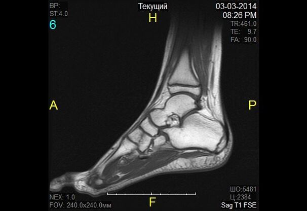
At nuclear magnetic resonance, changes in the properties of hydrogen molecules under the influence of strong magnetic fields are noted.
Magnetic resonance imaging
Magnetic resonance imaging (MRI) is an alternative diagnostic method that allows you to carefully examine the ligament structure of joint, muscle and cartilage tissues. With the help of an MRI, the doctor assesses the condition of the lower leg joints. Based on survey data, pathology is expressed in the early stages of development.
The diagnostic principle is based on exposure to radio waves and strong magnetic radiation. The magnetic field used is harmless and does not pose a health hazard.
MRI is contraindicated in case of mental disorders, during pregnancy and the presence of metal objects in the human body.
When diagnosing ankle arthrosis, a classic MRI machine (closed type) is used, as it has better picture quality. The MRI machine is a large cylindrical tube with a magnet around it. The patient lies on a special table. The ankle is fitted with a special coil. This procedure takes 30-40 minutes. This study was not painful at all. The patient may feel warmth in the lower leg area.
Ultrasound
Ultrasound examination has been widely used in medicine since the 90s of the 20th century. This technique has proven itself well in making an accurate diagnosis. Ultrasound scans are also performed for arthrosis of the ankle joint.
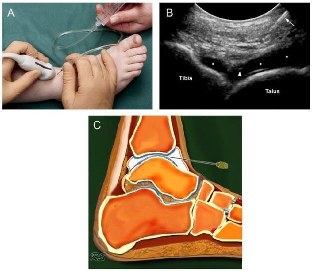
Today, ultrasound examination is not so important in the diagnosis of osteoarthritis, as it does not allow the study of damaged joints to be good enough.
The device conducting this study produces waves at ultrafrequency frequencies. The waves are reflected from the tissue and recorded on a monitor. Based on the resulting image, the doctor determines the type of pathology. To make the picture on the monitor clear, a special gel is used. This eliminates air gaps and gives the sensor a better glide.
Ultrasound examination does not harm the patient, so the procedure can be repeated many times. The advantages of ultrasound also include low cost and high accuracy.
The following indications are obvious signs of arthrosis:
- cartilage thinning;
- the presence of bone growth;
- accumulation of effusions in the joint cavity (synovitis);
- loss of cartilage space.
Bone scintigraphy
Scintigraphy is a high -precision study that, by using isotopes, can detect pathological changes in bone. Doctors divide the pathogen focus into "cold" and "hot". In the first case, we are talking about a zone where there are no isotopes. This area is deficient in blood, and cannot be seen during the scan. The "cold" area is the place where the malignant tumor is affected. In "hot" areas, isotopes quickly accumulate, and appear very bright when scanned. Such areas indicate the presence of inflammatory processes.
The role of scintigraphy in arthrosis is very significant. This study helps distinguish arthrosis from a number of other diseases when its clinical symptoms are similar.
During bone scintigraphy, a special preparation containing specially labeled atoms is injected into the body.
Based on the results of scintigraphy, the doctor makes a clinical prognosis and determines the treatment regimen. The only drawback of the study is its high cost. Scintigraphy is performed using special equipment, and, unfortunately, not all medical institutions can afford it.
Although radioactive scanning is a safe procedure, it still has some contraindications:
- pregnancy;
- lactation period;
- taking medications that contain barium.
When radioactive material is injected, some patients experience allergic reactions in the form of itching and rash. These side effects cause no harm and disappear on their own in a short time.
Joint puncture
Joint puncture is a diagnostic procedure in which a needle is inserted into an articular cavity to collect synovial fluid. This fluid is then sent for further research. Based on the data obtained, doctors draw conclusions about the nature of the disease and its stage of development.
At first glance, puncture is a simple procedure, but it is not. Fluid intake from the articular capsule requires exceptional physician movement accuracy. The synovium is very thin and an awkward movement of trauma. As a result, the inflammatory process develops. Potential risks also include infection. It is not difficult to bring the infection to the joint capsule through a device that is not properly sterilized.
The manipulation technique is different for each joint. While collecting articular exudate from the ankle, a puncture is made anteriorly, between the external ankle and the longus digitorum extensor tendon.
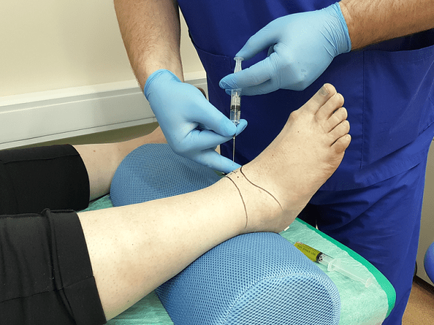
Diagnostic sampling of intra-articular fluid allows laboratory analysis and excludes arthritis.
Basic principles of treatment
After confirming the diagnosis of arthrosis of the ankle joint, the symptoms will not be long. Treatment is started immediately. Further prognosis depends on a well -chosen treatment regimen and the timeliness of initiation.
Arthrosis is a dangerous disease. It cannot be completely cured. The goal of therapy is to stop the degenerative process and prolong the remission period. For this purpose, doctors prescribe medications, physiotherapy, massage, rehabilitation gymnastics and folk remedies. If all conditions are met, it is possible to rely on positive dynamics, otherwise the disease continues.
Drug therapy for arthrosis
Depending on the effect of therapy, drugs are divided into several groups:
- Anti-inflammatory or painkiller. This group of drugs aims to eliminate the focus of inflammation and relieve pain. The earlier anti-inflammatory therapy is started, the more likely it is to save the joint. Drugs in this group can be produced in the form of tablets and ointments.
- Glucocorticoids. This drug is prescribed when the above funds are ineffective. They are produced in the form of solutions for injection. The medicine is injected directly into the joint.
- Chondroprotectors. Designed to slow cartilage destruction.
The regimen and dose of treatment is chosen by the doctor, based on the severity of symptoms, the age of the patient, the presence of concomitant diseases and other factors. Medications themselves are dangerous and often exacerbate the condition, as many medications have a number of side effects and have their own contraindications.

Features of radical treatment
If conservative therapy fails, then doctors are forced to use radical treatment methods (surgical intervention). Operations are also shown when:
- secondary (post-trauma) and primary arthrosis 3-4 degrees;
- arthrosis with complications;
- persistent and severe pain in the ankle, radiating to the knee;
- severe lameness;
- paresis and paralysis of leg muscles;
- violation of the flexion-extensor function of the joints;
- violation of leg support ability.
Surgical intervention is contraindicated if:
- patients under 12 years of age;
- fistulas are found in the joints;
- patients have a history of diabetes mellitus, heart failure;
- Infectious diseases were found in the proposed intervention area.
Traditional treatment
Doctors believe that the treatment of arthrosis should be carried out exclusively under the supervision of specialists, but they do not rule out the positive effects of folk remedies. Alternative medicine acts as an effective prophylaxis that helps relieve symptoms and maintain remission.
Folk remedies are a symptomatic treatment for leg arthrosis.
Home treatment should be coordinated with your doctor to avoid side effects and complications.
Traditional healers recommend treating ankle arthrosis by:
- Burdock. Wash burdock leaves with soap and running water. Apply the leaves with a gentle part to your skin. Fix the top with a bandage or adhesive film. It is better to keep the compress overnight.
- Sea salt. Cut the salt in the pan. Pour into a linen bag and attach to your ankles. Hold the bag until the salt is cool. Heat relieves pain. Sand, lentils, buckwheat are also used instead of salt.
- Lilac. Pour cologne three over the purple flowers. Leave the color in a dark, cool place for 10-14 days. Rub the affected area in the morning and evening.
- Eggshell. Grind the shells in a coffee grinder. Take the resulting powder for ½ tsp. before eat.
Do not forget that treatment with folk remedies should not be the only step. Complex treatments include medication, exercise therapy, massage, physiotherapy, spa treatments. In further cases, doctors use a radical measure - surgical intervention.
Surgery
For leg arthrosis, the following types of surgery are used in medicine:
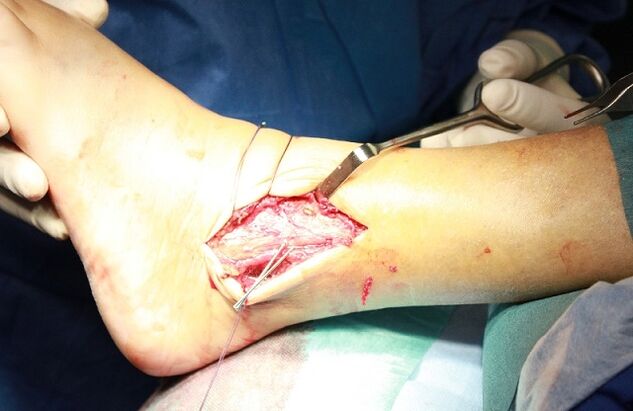
- arthrodesis of the joints;
- joint arthroscopy;
- endoprosthetics.
Arthrodesis is an operation to paralyze a joint. It is done to restore members lost support capabilities. The only drawback of the surgery is that the bones (tibia and talus) grow together, leading to a state of immobility. Arthrodesis is rarely used in medical practice.
Arthroscopy is a minimally invasive procedure. During the operation, the doctor makes a small incision in the joint area and through it inserts an arthroscope (a special tube at the end of the camera is fitted). With its help, the surgeon carefully examines and assesses the condition of the intra-articular structure. If necessary, a cut of the damaged joint or blood clot is removed from the synovial fluid. This manipulation is less traumatic. The only drawback of arthroscopy is that the risk of recurrence is too high.
Endoprosthetics are the last resort. It is performed with advanced arthrosis. Endoprosthetics allow you to replace an affected joint in part or in whole. As a prosthetic product, innovative prostheses with modern mechanics are used. Artificial joints last from 10 to 20 years.
Power characteristics
To achieve good results, drug treatment is supplemented with dietary therapy. Nutritionists have created a special diet to prevent worsening diseases and at the same time give the body all the necessary vitamins and nutrients. Diet for overweight patients plays a special role. Since obesity is one of the reasons for the development of arthrosis, weight correction is an important part of treatment.

The patient needs to reconsider some of his habits in daily life, which contribute to and provoke the development of leg arthrosis.
Nutritionists recommend adhering to the following dietary requirements:
- Eat regularly and in small portions.
- Drink at least 2 liters of fluid a day.
- Give candy and salt.
- The last meal is no later than 18: 00.
- Dishes are allowed to be steamed, boiled or baked.
The main task of the diet for arthrosis is a balanced and fortified diet. Fasting should not be questioned. Hard diets and body cleansing are more harmful than good. Calcium is excreted from the body, which is needed for cartilage recovery. A nutritionist will help you organize your daily diet.
With arthrosis, it is allowed to eat cereals, pasta, dairy products, cheese, legumes, vegetables, fruits, rye bread, dried fruit, nuts, fish, poultry meat. Heavy and fatty side dishes, foods containing dyes and flavors, as well as pickles, marinades, smoked meats, fatty broths, baked goods, spices, sauces, chocolate, ice cream, coffee and alcohol are prohibited.
Prevention of arthrosis
To prevent the development of arthrosis of the ankle joint, doctors recommend taking preventive measures:
- wear comfortable shoes without heels;
- stick to a diet and drink enough fluids;
- taking vitamin and mineral complexes seasonally;
- swim;
- walk more in the fresh air;
- removes excess pressure on the feet;
- avoid hypothermia;
- checked by a doctor in a timely manner.
With existing arthrosis, it is recommended to improve lifestyle:
- To push away from bad habits. It has been shown that they provoke blood stasis in tissues and accelerate the destruction of cartilage.
- Perform a set of exercises to warm up the ankles.
Prediction
Arthrosis is a progressive disease. Without treatment, it leads to irreversible consequences and complete joint instability. Early diagnosis of pathology allows you to do without radical measures. Medication can delay the pathological process and alleviate the patient's condition. The fight against the disease at an early stage without complications.